I started doing something a few years ago in preparation for my plastic surgery boards that triggered an epiphany about what humans find interesting. Here’s some background. For any plastic surgeon to become board certified, they must “collect” cases for approximately half a year after they pass their written board examination. This means that they keep a record of every operation they perform and take before, during and after-surgery photos with the patient’s knowledge. Additionally, they will collect any relevant laboratory or radiology tests and/or pathology results.
Eventually, the American Board of Plastic Surgery reviews all of these collected cases for each plastic surgeon that has passed their written boards but is waiting to take their oral boards. The Board chooses five cases that they want to review with the plastic surgeon. They ask to review specific cases, not because the doctor did something wrong or because there was a complication, but rather to ensure the board-eligible plastic surgeon is considering all of the signs and symptoms a patient exhibits and then making informed decisions based on those signs. And the Board isn’t just checking how well you answer their questions. They also give you very specific instructions on how to prepare the binder in which you place your chosen 5 cases. If you don’t follow their organizational instructions, they’ll send you home even before the test! But if you pass, you become a full-fledged board certified plastic surgeon. So where was I going with this? Oh, yes.
Part of the preparation process includes taking photos of every patient before and after surgery. And I got into the habit of taking photos during surgery to show the various stages of the operation. Say for example a patient came to me for removal of a cancerous mole on the forehead. I would photo-document what the mole looked like before surgery, the hole in the forehead after the cancer was removed, the flap of tissue or skin graft used to fill the hole and the final reconstructed result. This quickly evolved into taking pictures of everything I did during surgery. Since this was all digital photography, I wasn’t worried about the time and cost associated with developing film. I figured it would be safer to take more pictures than to take too few.
 As I did more cosmetic cases, my routine of taking photos of any intraoperative pathology specimen evolved into taking photos of the skin I removed for a tummy tuck or the fat I removed during a liposuction procedure. One day, a postoperative patient asked how much skin I removed. I didn’t weigh the skin so I couldn’t tell them exactly “how much” but I realized I could show them. I asked if they would like to see a photo and they very excitedly nodded their head. Fascinated is how I would describe their reaction. As any expert in a particular field can attest, we quickly forget that our floor may be someone else’s ceiling! While I may not think much of large swaths of skin or buckets of fat anymore, others, actually most everyone else, does.
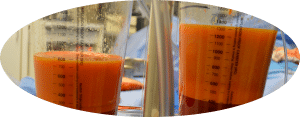
 So now I continue to take photos of everything I remove from the patient, even if it’s not required to go to the pathology lab and especially if it’s a cosmetic case with lots of skin and fat. The patients love to see it. Even if they initially think the idea of looking at their extra parts is disgusting, they immediately look at the photos when given the opportunity, while still claiming their disinterest!
Click here for the original blog post written by Dr. Jonathan Kaplan for BuildMyBod.?